Mammography (Mammogram)
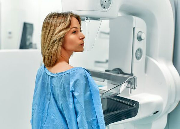
Mammography, which uses low-dose X-rays to create images, is the most basic method of breast imaging. Mammography is the method that can detect breast cancer before all other imaging and diagnostic methods. It can be described as an X-ray machine that can take images of the breast.
The purpose of mammography is to detect breast cancer early; the earlier it is detected, the more successful the treatment. Today, in addition to standard mammography methods, services are available using a variety of mammography technologies, most notably digital mammography.
Mammography Procedure
A mammogram requires some compression of the breast between the image sensor and the compression plate to produce an image. A standard mammogram takes two different images of each breast, one from the front and one from the side. If there are suspicious findings, additional images may be needed to better visualize and diagnose the area.
Why is compression necessary during a mammogram?
- To prevent breast displacement and obtain high-quality images
- Reduce breast thickness to lower the X-ray exposure.
- To expand overlapping tissue in the breast to avoid potential diagnostic errors.
How to Prepare for a Mammogram?
In such cases, patients often do not feel pain, only slight pressure. With typical chest pain, discomfort can be minimized by scheduling an examination in the second week of the period, that is, the week after the bleeding stops.
Cosmetics such as powder, deodorant, and shimmer cream should not be applied to the breast and armpit area during a mammogram. These substances can cause false and suspicious images and lead to diagnostic errors.