EEG
An EEG (electroencephalogram) is the process of recording the electrical activity of the brain, which is performed according to certain internationally accepted standards on digital media or paper-printed mappings. The brain is constantly electrically active, and this electrical activity shows significant levels of growth during certain periods of life and continues in specific patterns during certain phases of daily life (e.g., during sleep and waking).
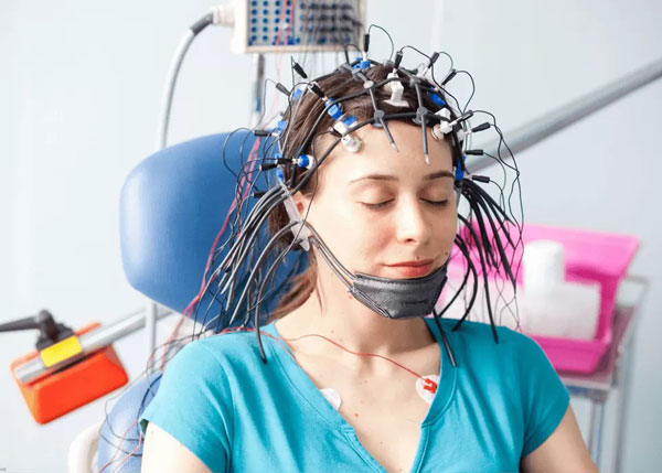
For a successful evaluation of an electroencephalogram, there are certain rules that need to be followed both by the technician and the patient while recording the brain waves on the computer equipment.
To record the patient’s brain waves, as a standard procedure, electrodes are placed on the scalp and earlobes for the purpose of amplifying the signals.
The essential steps to be taken during the recording process are as follows:
- One monopolar electrode on the earlobe and the other electrode on the scalp
- Bipolar recording with connections on the scalp of both electrodes,
- Recording eye movements and
- Using seizure provocation methods.
Seizure provocation methods in routine EEG,
The use of hyperventilation during intermittent stimulation by light at different frequencies.
The seizure provocation can also be triggered by sound or, if necessary, by insulin, which is less commonly used today.
Sleep also has a special effect on the brain's electrical activity at different stages. In addition to spontaneous and electrically induced sleep EEG recordings, we can offer other equipment (eye movement, electrocardiogram, electroencephalogram, and simultaneous recording of breathing) to record all stages of sleep and investigate sleep disorders, sleep paralysis, and sleep epilepsy (polysomnography).
In cases where no pathological findings are found in the interictal periods between seizures, telemetric EEG monitoring and long-term EEG monitoring can be performed for recording in daily life, and simultaneous recording with video for recording the clinical seizure with any changes in the EEG.
Important factors to consider when performing an EEG
Age, gender, preliminary diagnosis, clinical information, medications and dosages, patient's condition at the time of recording (anxiety, drowsiness, coma, diazepam administration, etc.), cooperation, and relationship to the environment should be indicated. In addition, eye-opening and closing, photostimulation and hyperventilation, and notable conditions (swallowing, lying down, seizures, etc.) should be marked on the footprints simultaneously for the most accurate assessment.
The assessment may then proceed and the next steps may follow in order.
Is the baseline activity consistent with the patient's age, current condition, medication and disease? Is there a significant difference from previous recordings? (the organization of baseline activity may not be normal in certain metabolic, inflammatory, degenerative diseases, which show certain characteristics in certain age groups, drug use and sleep patterns).
The effects of opening or closing eyes (eyes-open state mostly causes brainwave suppression)
Presence or absence of asymmetry between the two hemispheres of the brain (subdural hematoma, cerebral infarction, mass can sometimes cause suppression and delay, unilateral encephalitis can cause asymmetry in the form of delay).
Presence or absence of focal and multifocal abnormalities (structural, degenerative, metabolic, metabolic, inflammatory, vascular and neurocutaneous causes)
The presence or absence of the paroxysmal abnormality, its spread, origin, outcome and type, if any (it may occur during epilepsy and is useful for epilepsy criteria, if obtained).
The effect of intermittent photostimulation (necessary to investigate the presence of photosensitive activities) and hyperventilation on the EEG trace will be evaluated and is useful for the clinical and electrophysiological detection of certain cases of epilepsy.
Sleep EEG is performed in infants and uncooperative children, and although it uses the same assessment criteria, except for hyperventilation and open-eye state, understanding the infant EEG and sleep EEG criteria for proper assessment will minimize false abnormal or false normal findings.
For what purpose is an EEG performed?
EEG is mainly used to conduct research on epilepsy. However, because seizures are not always clinically recorded and the duration of EEG recording is limited, even if the patient is epileptic, abnormal findings may not be detected on the EEG during the inter-seizure phase and this does not mean that the patient is not epileptic.
On the contrary, the detection of epileptiform activations on an EEG in a patient without clinical seizures does not necessarily indicate the presence of epilepsy.
The EEG reaches significance in cases of epilepsy when clinical and EEG findings coincide and are essential in monitoring patients with a diagnosis of epilepsy. In addition, in cases of epilepsy, the detection of seizures on EEG will be useful in determining case criteria and treatment options.
In epileptic cases, focal, multifocal or asymmetric findings in the intraoperative period may be an indication of epileptic foci, requiring the exclusion of structural pathology.
Infections (herpetic encephalitis, subacute sclerosing pancreatitis, Jacob Kreutzfeldt's disease, etc.), toxic, metabolic (hypoglycaemia, hyperglycaemia, hepatic encephalopathy, etc.), degenerative, hypoxic (heart and lung failure, CO poisoning) encephalopathies, electrical modulation of the brain can help in diagnosis and prognosis.
EEG can be used in the differential diagnosis of certain behavioral disorders, enuresis and syncope, in unexplained and refractory, recurrent clinical conditions (refractory tachycardia attacks, abdominal pain attacks, infantile spasms mimicking abdominal pain, etc.), in the differential diagnosis of epileptic phenomena and the investigation of sleep disorders.