Echocardiography
Echocardiography is a study of the structure and function of the heart using sound waves (ultrasound). It is performed with a device (transducer) that emits sound waves. The transducer is attached to different parts of the chest and the walls and valves of the heart are examined at different locations.
There are 4 basic methods of echocardiography.
- Transthoracic (Surface) Echocardiography (TTE)
- Transoesophageal echocardiography (TOE)
- Stress echocardiography
- Strain and Strain Rate Imaging by Echocardiography
- How is Transthoracic (Surface) Echocardiography
(TTE) performed?
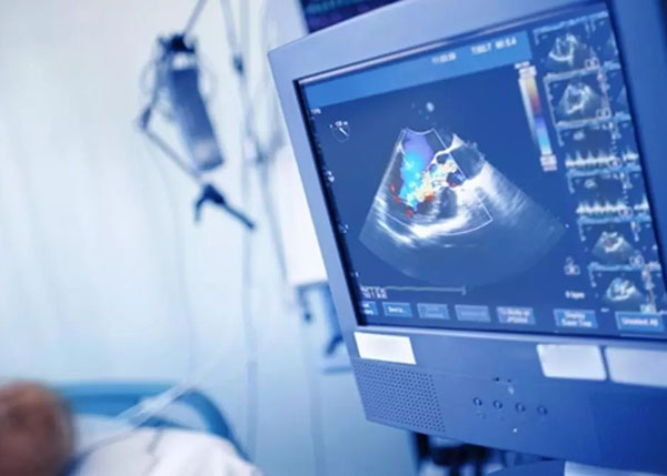
In Transthoracic (Surface) Echocardiography, the patient is positioned on the left side and a gel transducer on the patient's chest is moved across to examine different parts of the heart. X-rays are not used in this test. In reality, Transthoracic (Surface) Echocardiography is an ultrasound technique. The examination is usually completed in 15 minutes. It is a rather painless procedure and has no side effects. The screen shows cross-sections and movements of the heart valves, and large vessels (aorta, pulmonary artery). The results are evaluated by the physician performing the exam.
Why might you need Transthoracic (Surface) Echocardiography?
- Detect the cause of any unusual sounds in the heart (such as a heart murmur) heard with a stethoscope,
- Search for the cause of an enlarged heart, unexplained chest pain, shortness of breath, or irregular heartbeat,
- Measure the shape and size of the heart chambers
- Check the thickness and movement of the walls of the heart,
- Clearly assess the structure and movement of the heart valves,
- Evaluate the function of a prosthetic heart valve,
- Evaluate heart function,
- Detect diseases that affect the heart muscle (such as cardiomyopathy)
- Evaluation of blood clots and tumors in the heart,
- To check for congenital heart disease or surgery performed because of it,
- To evaluate heart function after a heart attack,
- If there is fluid buildup around the heart, assess the amount and type of fluid and evaluate the structure and thickness of the pericardial membrane that surrounds the heart,
- To evaluate the structure and diameter of the major arteries that leave the heart (aorta, pulmonary arteries).
What is Transoesophageal echocardiography (TOE)?
In certain cases, when the usual surface screening method is not satisfactory, a transoesophageal screening may be requested. This procedure is akin to a gastroscope. Because transoesophageal echocardiography is also performed through the esophagus.
Why might you need Transoesophageal echocardiography?
- For further examination of diseases detected by surface echocardiography such as clots, masses, or endocarditis due to infection inside the heart,
- To analyze the functions of artificial valves in detail,
- When suspected aortic vessel dilatations and ruptures,
- To analyze the holes in the membranes between the chambers of the heart,
- Determining the severity of valvular heart failure,
- Evaluation of surgical success during and after heart valve repair or closure of heart holes,
- When surface echocardiographic images of high resolution cannot be obtained due to lung disease, obesity, or chest structure, the TOE method is used.
How to prepare the patient before Transoesophageal echocardiography?
Transesophageal echocardiography is performed after a fast of 4-12 hours. Patients with allergies, asthma, increased eye pressure, difficulty swallowing, nasal congestion, new throat infections, and esophageal and stomach problems should inform the doctor running the test.
How is Transoesophageal echocardiography performed?
Transoesophageal echocardiography is a semi-interventional test. Immediately before the examination, intravenous access is established so that medications can be administered as needed. The mouth and soft palate are locally anesthetized with an anesthetic spray to suppress the gag reflex and ensure patient compliance.
Intravenous sedation is administered for the purpose of the procedure and for patient comfort. If the patient does not comply, the examination is resumed with further sedation under the supervision of an anesthesiologist.
The cardiologist will explain to the patient how to swallow the tube. A mouthpiece will be placed in the mouth to prevent the patient from chewing the tube. The gel-lubricated transesophageal echocardiography probe is slowly inserted through the esophagus. If the patient swallows the tube, vomiting and nausea are normal. It is temporary. During this time, breathe in and out through the nose.
If necessary, the physician will videotape and take pictures of the heart. After the examination, the physician will inform you about the findings. The average examination time is 15-20 minutes. However, with preparation time, this time can reach 30-60 minutes.
After the Transoesophageal echocardiography procedure
After the procedure, you must not eat or drink anything for about 2 hours until the numbness in your throat is completely gone. As the medicines used to calm you down during the examination may cause drowsiness for a period of time, you should not drive or use machinery until this is completely reversed. After the procedure, you may experience sore throat and loss of feeling for 1 to 2 days. This is a temporary condition that does not require treatment.
What is stress echocardiography?
Stress echocardiography (SE) is a test performed with exercise methods or medicine that increase heart rate. In exercise echocardiography, echocardiographic images are recorded immediately before and after an exercise protocol in a treadmill stress test or at each exercise step in a bicycle stress test. If an exercise stress test cannot be performed (vascular disease of the lower extremities, muscular or skeletal limitations), drug echocardiography is performed with intravenous drugs such as dobutamine, adenosine, or dipyridamole administered in increasing doses at regular intervals to increase heart rate and contraction.
Why might you need stress echocardiography?
Permanent cardiac pacemakers are used when left bundle branch block, left ventricular thickening and certain specific electrocardiographic findings (pre-excitation) make it difficult to assess cardiac disease with other methods. In particular, it is applied to determine myocardial blood flow abnormalities and their severity, to identify risk after acute myocardial infarction and coronary intervention procedures, and to grade preoperative cardiac safety in patients undergoing other than cardiac surgery.
The main purpose is to observe the contractility of the heart, to look for any signs of coronary artery disease, and to help in making surgical decision-making for certain heart valve diseases.
How to prepare for stress echocardiography?
Stress echocardiography requires fasting for an average of 4-6 hours. During this 6-hour period, smoking should be avoided and caffeine-containing foods (tea, coffee, chocolate, colas, etc.) and medications (because some painkillers contain caffeine) should not be consumed.
Patients should stop taking certain medications 24 hours prior to the test (to be determined by the physician ordering the test). Approved medications may be swallowed with a small amount of water 3-4 hours prior to testing.
For all tests requiring fasting, patients with diabetes should not take any blood glucose control medications before a meal is allowed. Meals may be consumed immediately following the SE examination.
How is stress echocardiography performed?
Preparation for the test consists of attaching electrodes to your chest and opening the establish vascular access. The test takes about an hour to complete. The test is performed by recording at specific points on your chest. An image of your heart at rest is recorded.
Depending on your preferred stress test, a physical exercise will be performed or medication will be administered. Images are taken during physical activity and then recorded during the recovery period. Your heart rate, blood pressure, and electrocardiogram will be monitored.
During the test, fast and strong heartbeats will be recognized as heart palpitations. During a drug test (dobutamine injection), symptoms such as warmth and redness of the cheeks, and tingling of the scalp are normal. If during the procedure you experience pain and discomfort in the chest, arms, jaw, dizziness, darkening of the eyes, and difficulty breathing, you should immediately inform the doctor who performed the procedure.
The patient is monitored in the recovery room for 30 minutes after the procedure. The results of the examination are interpreted by comparing the intensity of the heartbeat in the images taken at different stages.
The results of stress echocardiography are explained to the patient by the doctor and a written report is provided to the patient within an hour.
Does exercise echocardiography pose any risks?
Stress echocardiography is a completely safe procedure. Drug-related adverse effects are very rare to occur with stress echocardiography. These may include headache, sweating, palpitations, chest pain, shortness of breath, and nausea.
In uncommon cases, sudden drops and increases in blood pressure, sudden and non-sustained ventricular tachycardia arising from the atria and ventricles, decreased heart rate, and chest pain of cardiovascular origin may occur during the procedure.
What is Strain and Strain Rate Imaging by Echocardiography?
Strain and Strain Rate Imaging by Echocardiography is the most accurate, newest, and cutting-edge method designed to precisely measure the contractile and relaxation functions of the heart in different areas using tissue Doppler ultrasonography. This method is used in particular to evaluate the strength and contractile function of the heart muscle. Different areas of the heart muscle are examined, and their dimensions and movements under stress and at rest are monitored in different areas.
The procedure is completely painless and has no side effects. No preliminary preparation is to be necessary before the procedure. No contrast agent is used. The procedure takes an average of 15-30 minutes.
The results are both evaluated by the examining physician and shared with the patient. After the procedure, the patient can get up immediately and resume daily life.
Why might you need Strain and Strain Rate Imaging by Echocardiography?
The strength of the contraction of the heart muscle in a patient with heart failure is most precisely measured. This allows the cause of heart failure to be detected much earlier than the normal eye can see and the most appropriate treatment option to be determined.
For a patient who suffered a heart attack, this method takes very precise accurate measurements to determine the mass of the damaged heart muscle and the extent of the damage. It is one of the main methods used to assess heart muscle function before any heart surgery. It is the most reliable way to detect pericardial inflammation and distinguish between its types.
It is a unique method for the immediate detection of simultaneous dysfunction of the right and left halves of the heart, as well as for determining the treatment used and its effectiveness.